Chest Radiography Stand — Practical Guide for Buyers & Clinicians
What is a chest radiography stand?
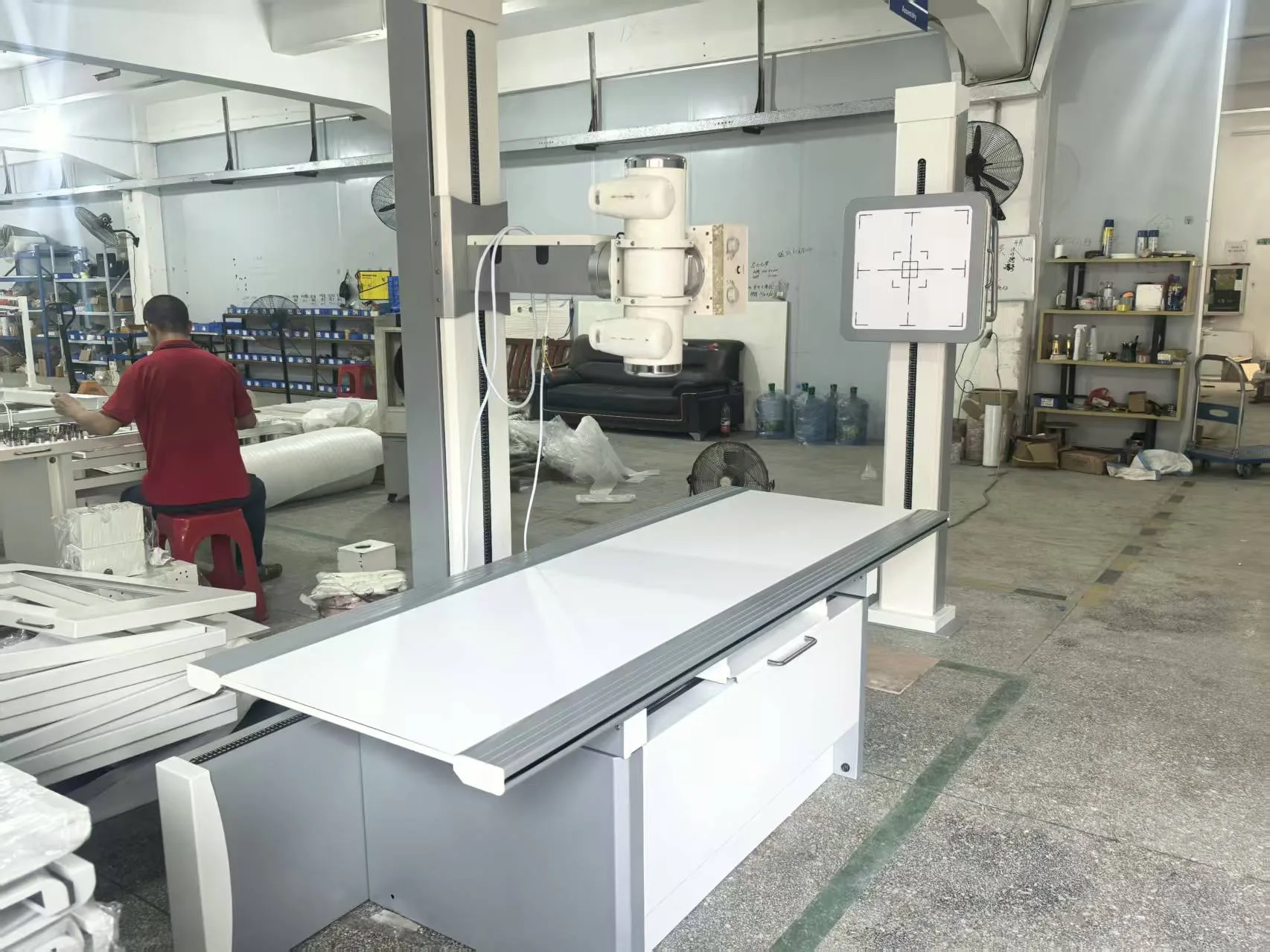
A chest radiography stand is a stable, vertically adjustable frame that holds the image receptor — whether a film cassette or a flat panel detector — in front of the patient during upright X‑ray exams. Originating as a standard accessory after X‑ray grew common in the early 20th century, the stand continues to be an essential element in outpatient and inpatient thoracic imaging.
Key components and operation
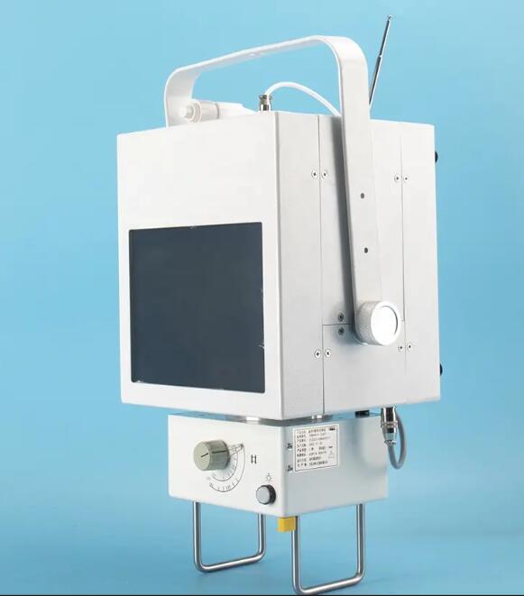
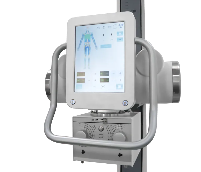
Typical stands include a weighted base, a vertical column, and a receptor holder with height and tilt adjustment. Modern models offer smooth manual gearing or electric lifting, quick-release clamps for detector insertion, and angle scales for reproducible positioning. The goal is simple: align the X‑ray beam, patient chest, and receptor so the image is sharp and consistently reproducible.
- Base & column: stable geometry to avoid drift and vibration.
- Receptor mount: compatible with cassettes and flat panels.
- Height & angle control: manual, geared, or electric drive options.
- Locking mechanisms: secure positioning to reduce retakes.
Clinical and workflow benefits
A well‑designed chest stand improves image quality, reduces repeat examinations, and shortens room turnaround time. Key advantages include:
- Height adaptability for different patient statures and bed heights.
- Reliable positioning that lowers retake rates and radiation exposure.
- Faster exam setup when combined with clear scales and intuitive handles.
- Compatibility with digital detectors enables quick integration with PACS.
How to choose the right stand — Buyer checklist
When selecting a chest radiography stand for a clinic, hospital or export shipment, check the following criteria:
- Detector compatibility: mechanical fit and cable routing for your flat panels or cassettes.
- Adjustability: travel range, smoothness of lift and repeatable locking positions.
- Build quality: robust base and corrosion-resistant materials for continuous clinical use.
- Ergonomics: handle placement, scales, and ease of cleaning.
- Safety & compliance: shielding, electrical safety, and relevant certifications (CE, FDA or local approvals where applicable).
- Service & spares: local support, spare parts availability and training for staff.
Installation, maintenance and safety tips
Install the stand on a level surface and verify locking mechanisms before first clinical use. Periodic checks should include lubrication of moving parts, inspection of electrical wiring (for motorized models), and verification of detector mounting integrity. Maintain logs for preventive maintenance and train staff on correct patient positioning to avoid unnecessary exposures.
- Height range: 900–1800 mm
- Detector compatibility: 14″x17″ / 17″x17″ flat panels
- Lift: electric (24 V) or manual with worm gear
- Finish: anti-corrosive powder coat
- Certifications: CE, ISO 13485 (examples)
Final considerations
The chest radiography stand is a deceptively simple but crucial component of thoracic imaging. Selecting a model that fits your detectors, clinical workflows and maintenance capacity will pay back quickly in reduced retakes and faster exams. For exporters, emphasize documented compliance, spare-part kits and clear installation guides to help international buyers integrate the stand into their radiology suites.