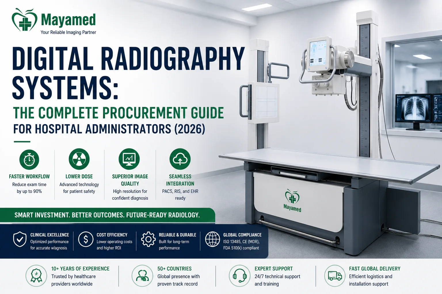
Digital Radiography Systems: The Complete Procurement Guide for Hospital Administrators (2026)
Article Summary
The Evolution of digital radiography: Why Upgrade in 2026? The global digital radiography market is projected to reach $6.
The Evolution of digital radiography: Why Upgrade in 2026?
The global digital radiography market is projected to reach $6.8 billion by 2028, driven by a robust 5.4% CAGR. This growth is not merely a reflection of technological maturation; it is a direct response to the urgent clinical and operational mandates facing modern healthcare facilities. As emerging markets rapidly transition from Computed Radiography (CR) to Digital Radiography (DR), and established institutions replace aging legacy DR units, the strategic importance of selecting the right digital radiography system has never been higher.
For hospital administrators and radiology directors, the operational delta between CR and modern DR is staggering. Transitioning from legacy CR to DR reduces average image acquisition and processing time from 90 seconds to under 10 seconds. In a high-volume environment, this efficiency gain translates to an increase in daily patient throughput of up to 35%. Furthermore, the elimination of CR cassettes, imaging plates, and laser readers drastically reduces consumable costs and mechanical points of failure.
However, upgrading hospital radiology equipment in 2026 requires looking beyond simple image acquisition. Today’s procurement decisions must account for artificial intelligence integration, total cost of ownership (TCO), and seamless interoperability within complex Health Information Technology (HIT) ecosystems. This guide provides an end-to-end framework for evaluating, procuring, and deploying advanced radiography systems that balance clinical excellence with strict fiscal and infrastructural realities.
Core Components of a Modern Digital Radiography System
A high-performance digital x-ray machine is a symphony of precisely calibrated hardware and software. When evaluating configurations, procurement teams must assess each subsystem not in isolation, but for how it impacts overall workflow and diagnostic confidence.
High-Frequency X-Ray Generator
The generator is the powerhouse of the system. Modern systems utilize high-frequency (HF) generators operating between 50 kW and 80 kW. A higher kW rating allows for shorter exposure times, which is critical for minimizing motion artifacts in trauma, pediatric, and uncooperative patients. Look for generators capable of delivering consistent high-voltage waveforms with less than 1% ripple, ensuring optimal X-ray tube output and dose efficiency.
X-Ray Tube and Collimator
The tube’s heat capacity (measured in MHU or kHU) dictates how many consecutive exposures can be taken before thermal cooldown is required. For busy emergency departments, a tube with a minimum of 300 kHU is recommended. Integrated motorized collimators with auto-beam limiting are essential for dose optimization and workflow speed.
The flat panel detector (FPD)
The flat panel detector is the most critical and expensive component of the imaging chain. Modern systems utilize either wired or wireless FPDs. Wireless detectors with integrated batteries and Wi-Fi/Bluetooth telemetry significantly enhance patient throughput by allowing technologists to position the detector without being tethered to the bucky or wall stand.
Acquisition Workstation and IT Integration
The workstation must feature high-resolution, medical-grade diagnostic monitors (minimum 3MP for general radiography, 5MP for mammography/tomosynthesis). More importantly, the software architecture must natively support DICOM 3.0, HL7, and IHE (Integrating the Healthcare Enterprise) profiles to ensure seamless communication with your existing PACS, RIS, and EHR.
Flat Panel Detector Technology: CsI vs. a-Se and Impact on Dose
Understanding the physics of the detector is non-negotiable for informed procurement. The market is dominated by two primary scintillator technologies: indirect conversion using Cesium Iodide (CsI) and direct conversion using Amorphous Selenium (a-Se).
Cesium Iodide (CsI) scintillator flat panel detectors maintain approximately 78% market share in general radiography. This dominance is due to their superior Detective Quantum Efficiency (DQE) at lower mAs settings. CsI is grown in structured needle-like crystals that channel light photons directly to the photodiode array, minimizing lateral light spread and preserving spatial resolution while maximizing X-ray absorption.
Conversely, a-Se detectors absorb X-rays directly and convert them into electrical charge, eliminating the light-scattering step entirely. While a-Se offers exceptional intrinsic spatial resolution (ideal for extremity imaging), its DQE drops off more rapidly at higher spatial frequencies compared to structured CsI, making CsI the preferred choice for general chest and abdominal radiography where contrast resolution and low-dose performance are paramount.
Industry Insight: "From a biomedical engineering perspective, the shift to solid-state FPDs has been a revelation. Legacy CR readers were plagued by mechanical failures—laser scanners, transport rollers, and erasure lamps required constant preventive maintenance. Modern FPDs have virtually no moving parts. The hidden maintenance costs and downtime associated with CR readers have been entirely eliminated, shifting our operational focus from reactive hardware repair to proactive software and network management."
— Director of Biomedical Engineering, Regional Health Network
Key Technical Specifications to Evaluate: DQE, Generator Power, and Spatial Resolution
Vendor specification sheets are often dense with marketing jargon. Procurement managers and clinical leads must filter this data to focus on the parameters that directly impact image quality and patient dose.
| Technical Parameter | Clinical / Operational Impact | Target Specification for General Radiography |
|---|---|---|
| DQE (0 lp/mm) | Overall dose efficiency. Higher DQE means lower patient dose for the same image noise level. | ≥ 65% - 70% at 70 kVp |
| DQE (2 lp/mm) | Contrast resolution at higher spatial frequencies. Critical for visualizing fine trabecular bone and lung markings. | ≥ 35% - 40% at 70 kVp |
| Pixel Pitch | Determines maximum spatial resolution (Nyquist frequency). Smaller pitch = finer detail, but larger file sizes. | 139 µm or 143 µm (Standard); 100 µm (Extremity) |
| Generator Power (kW) | Dictates minimum exposure time. Crucial for freezing patient motion. | 65 kW (Standard); 80 kW (High-volume/Trauma) |
| kVp Range | Penetrating power. Must cover pediatric (40 kVp) to large lateral lumbar (125-150 kVp). | 40 to 150 kVp |
When evaluating the digital radiography system, request raw DQE curves measured according to the IEC 62220-1 standard. Do not rely solely on the peak DQE at 0 lp/mm; examine the curve's drop-off at 1 and 2 line pairs per millimeter (lp/mm) to ensure the detector maintains contrast resolution for subtle pathologies.
Total Cost of Ownership (TCO) vs. Initial dr system Price
The initial DR system price is only the tip of the financial iceberg. A rigorous TCO model is essential for accurate capital planning. Average DR system prices range from $120,000 for basic mobile/portable units to over $450,000 for advanced ceiling-suspended U-arm systems with dual wireless detectors and AI software packages.
Deconstructing the TCO
- Consumables and Accessories: While DR eliminates CR plates, it introduces costs for detector protective covers, grid inserts, and specialized anatomical positioning aids.
- X-Ray Tube Replacements: An X-ray tube is a consumable component with a finite lifespan (typically 100,000 to 300,000 exposures). A tube replacement can cost between $8,000 and $15,000. Factor in the expected replacement cycle (usually years 5 to 7).
- Detector Battery Replacements: Wireless FPD batteries degrade over time. Budget for battery replacements every 3 to 4 years per detector.
- Service and Maintenance Contracts: Post-warranty comprehensive service agreements typically cost 6% to 10% of the original equipment cost annually.
- IT Integration and Cybersecurity: Costs for DICOM routing configuration, firewall exemptions, and ongoing cybersecurity patching for the acquisition workstation.
When comparing premium OEMs (such as Siemens Healthineers, GE HealthCare, and Philips) against value-tier manufacturers, the initial DR system price gap can be substantial. However, value-tier systems may carry higher long-term TCO due to shorter tube lifespans, lower DQE (resulting in higher retake rates), and less robust localized service networks. Always demand a 5-year or 7-year TCO projection from the vendor during the RFP process.
Navigating Medical Imaging Procurement: RFP, Vendor Selection, and SLAs
Effective medical imaging procurement requires a structured, cross-functional approach. The Request for Proposal (RFP) must be co-authored by radiology leadership, biomedical engineering, and IT to ensure all operational vectors are covered.
Critical RFP Components
- Clinical Workflow Requirements: Define daily exam volumes, required anatomical packages, and specific positioning needs (e.g., weight-bearing, trauma cross-table).
- IT and Interoperability: Specify required DICOM Storage, Worklist, and Modality Performed Procedure Step (MPPS) capabilities. Require a detailed Interface Control Document (ICD) timeline.
- Training and Application Support: Mandate a minimum of 40 hours of on-site clinical application training for technologists, plus dedicated training for biomedical engineers on first-line troubleshooting.
Negotiating Service Level Agreements (SLAs)
Do not accept boilerplate service contracts. Negotiate SLAs that include financial penalties for non-compliance. Key metrics should include:
- System Uptime: Guaranteed at 99.5% or higher, calculated monthly.
- Response Time: Maximum 4-hour on-site response for critical failures (system down), and 24-hour response for non-critical issues.
- Parts Availability: Guarantee that 95% of critical spare parts (detectors, generator boards) are stocked within a 100-mile radius of your facility.
Facility Preparation: Power, Shielding, and Structural Requirements
One of the most common pitfalls in hospital radiology equipment deployment is underestimating facility preparation. A delayed installation can cost tens of thousands of dollars in lost revenue and project overruns.
Structural and Spatial Requirements
Ceiling-suspended U-arm systems are the gold standard for high-volume rooms, but they impose massive structural loads. The ceiling track and U-arm suspension can weigh over 1,500 lbs. This requires structural engineering analysis to reinforce the ceiling joists or install dedicated steel unistrut frameworks tied to the building's primary load-bearing columns. Floor loading must also be assessed if utilizing floor-mounted systems.
Power Infrastructure and Emerging Market Challenges
High-frequency generators draw significant instantaneous power. A dedicated 480V/3-phase power line with a minimum 100-amp service is typically required. In emerging markets or older facilities with unstable power grids, voltage sags and frequency fluctuations can damage sensitive generator inverters and FPD electronics. Procurement must include the installation of heavy-duty online double-conversion UPS systems and automatic voltage regulators (AVRs) specifically sized for the X-ray generator's peak kVA demand.
Radiation Shielding
While the shift to DR often allows for lower patient doses, the shielding requirements are dictated by the workload (mAs/week) and the kVp utilized. Ensure your medical physicist has updated the shielding calculations based on the specific use factors of the new equipment, particularly if upgrading from a low-output CR system to a high-output 80kW DR system.
Regulatory Compliance and Certifications for Global and Emerging Markets
Procurement in the medical device sector is inextricably linked to regulatory compliance. Ensuring your equipment meets international standards mitigates legal risk and ensures compatibility with local health authority mandates.
- ISO 13485: Verify that the manufacturer’s Quality Management System is certified to ISO 13485, ensuring consistent design and manufacturing controls.
- CE MDR (Medical Device Regulation): For European and many global markets, ensure the device complies with the new EU MDR (2017/745), which has significantly stricter clinical evaluation and post-market surveillance requirements than the old MDD.
- FDA 510(k): For the US market, verify the specific model has a cleared 510(k) premarket notification. Do not rely on a "family clearance"; ensure the exact configuration is listed.
- IEC 60601-1 & IEC 60601-1-2: Confirm compliance with the general safety standard (60601-1) and the electromagnetic compatibility (EMC) standard (60601-1-2). EMC is critical to prevent the X-ray generator from interfering with adjacent life-support equipment.
- IAEA Guidelines: Align your dose optimization protocols with the International Atomic Energy Agency (IAEA) human health publications on diagnostic reference levels (DRLs).
Future-Proofing Your Radiology Department: AI Integration and Cloud PACS
The modern digital X-ray machine is no longer just an image capture device; it is an intelligent data node. Artificial intelligence is fundamentally altering radiography workflows, moving from post-processing analysis to real-time acquisition assistance.
AI-Assisted Auto-Collimation and Positioning
Advanced DR systems now feature AI algorithms that automatically detect anatomical landmarks and body contours in real-time on the acquisition monitor. This allows for auto-collimation, ensuring the X-ray beam is restricted precisely to the area of clinical interest.
Hospitals that integrate AI-assisted auto-collimation and positioning in their DR systems report an average 15-20% reduction in repeat imaging rates and associated radiation dose. By eliminating cone-cut errors and ensuring consistent anatomical coverage, AI directly improves first-pass yield.
Clinical Perspective: "The integration of AI-driven image quality enhancement and auto-exposure optimization has been a game-changer for our pediatric protocols. We are now able to utilize ultra-low dose techniques with confidence. The AI algorithms effectively suppress quantum mottle without blurring fine anatomical edges, allowing us to maintain strict ALARA (As Low As Reasonably Achievable) principles for neonates and infants without compromising the diagnostic confidence of our attending radiologists."
— Chief Radiologist, Children’s Specialty Hospital
Cloud PACS and Edge Computing
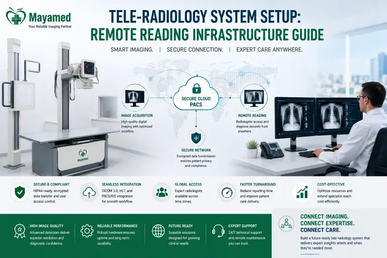
For multi-site health networks and emerging market tele-radiology hubs, evaluate systems that offer native edge computing and secure cloud PACS integration. This allows for immediate image offloading, enabling radiologists to read studies from remote locations in real-time, effectively decoupling image acquisition from image interpretation.
Real-World Deployment Scenarios
To contextualize these technical and financial parameters, consider how different configurations solve distinct operational challenges in real-world environments.
Case Study 1: High-Volume Urban Trauma Center
The Challenge: A Level 1 Trauma Center was experiencing severe workflow bottlenecks in the emergency department. The single-room DR setup, utilizing an older floor-mounted system with a single wired detector, could not keep pace with the 150+ daily trauma and acute care radiographs. Patient wait times were excessive, and door-to-image times were critically delayed.
The Solution: The facility procured a high-end, ceiling-suspended U-arm digital radiography system equipped with dual wireless flat panel detector panels (one 14x17 inch and one 10x12 inch) and an 80 kW high-frequency generator. The system was integrated with AI-driven auto-positioning software.
The Outcome: The dual-detector configuration eliminated the 45-second detector swap time. The ceiling-suspended U-arm allowed for rapid, multi-angle trauma cross-table imaging without moving the patient. Consequently, the department reduced door-to-image time by 40% and eliminated the radiography workflow bottleneck, increasing overall ED throughput by 22% without adding additional technologist headcount.
Case Study 2: Rural/Emerging Market Clinic
The Challenge: A rural clinic in an emerging market required diagnostic-grade imaging capabilities but faced severe infrastructure limitations. The facility had a highly unstable local power grid with frequent brownouts, limited physical space for a dedicated radiology room, and no access to a specialized biomedical engineering team.
The Solution: The clinic deployed a battery-operated, fully mobile DR unit. The system featured a high-DQE (68% at 0 lp/mm) wireless flat panel detector and an integrated, medical-grade UPS capable of sustaining 20 exposures on a single battery charge, independent of the local grid. The software included automated exposure control (AEC) calibrated for varying grid voltages.
The Outcome: The battery-operated mobile DR bypassed the need for expensive facility electrical upgrades. The high-DQE detector ensured diagnostic image quality was maintained even when operating on reduced battery voltage. The system's ruggedized design and solid-state FPD minimized the need for specialized maintenance, ensuring continuous operation in a resource-constrained environment.
Actionable Next Steps for Procurement Leaders
Procuring a new digital radiography system is a complex, multi-disciplinary endeavor. To ensure a successful deployment that maximizes ROI and clinical efficacy, execute the following steps immediately:
- Conduct a Baseline Workflow Audit: Quantify your current daily exam volumes, peak hours, and repeat rates. Use this data to justify the ROI of dual-detector or AI-enabled configurations.
- Initiate a 5-Year TCO Model: Move beyond the initial DR system price. Mandate that vendors provide detailed 5-year cost projections including tube replacements, battery swaps, software licensing, and comprehensive service contracts.
- Engage Facilities Engineering Early: Before finalizing the RFP, have your facilities team assess the structural load capacity for ceiling-suspended tracks and the electrical infrastructure for high-kW generators, particularly regarding voltage stabilization for unstable grids.
- Demand Clinical Trials: Do not purchase based on spec sheets alone. Require a 2-week on-site clinical trial with the exact detector and generator configuration proposed, allowing your technologists and radiologists to evaluate the workflow and image quality in your specific environment.
- Lock in SLA Penalties: Ensure your legal and biomedical teams negotiate ironclad Service Level Agreements with financial penalties for uptime failures and delayed response times.
Frequently Asked Questions
What are the specific facility infrastructure requirements, particularly regarding power supply and cooling, for installing this high-frequency digital radiography system?
Our DR systems are engineered with emerging market infrastructure in mind, featuring a wide voltage tolerance range and built-in power conditioning to handle unstable grid fluctuations without requiring expensive external voltage stabilizers. The X-ray tube and generator assembly utilize advanced oil-cooling and heat dissipation technologies, significantly reducing the need for dedicated, high-capacity HVAC systems in the scanning room. During the pre-installation phase, our biomedical engineering team conducts a comprehensive site survey to verify your facility's electrical grounding, power capacity, and ambient temperature controls, ensuring a seamless deployment without costly facility upgrades.
Does the system hold current FDA 510(k) clearance and CE marking, and how does the manufacturer ensure ongoing compliance with local radiation safety standards in emerging markets?
Yes, our digital radiography systems are fully FDA 510(k) cleared and carry the CE mark under the Medical Device Regulation, ensuring baseline global compliance for international procurement. Beyond these international certifications, we provide comprehensive technical dossiers and localized radiation safety documentation to assist your facility in obtaining approvals from local atomic energy or health ministries. Furthermore, our systems are equipped with integrated, tamper-proof dosimetry tracking and automated exposure control calibration tools that continuously monitor and log radiation output, guaranteeing ongoing adherence to ALARA principles and local regulatory audits. We also offer remote software updates to ensure your system's safety protocols are always aligned with the latest international radiation protection guidelines.
How does the preventive maintenance program and parts availability impact the Total Cost of Ownership (TCO) and system uptime over a standard seven-year lifecycle?
To minimize Total Cost of Ownership, we offer tiered comprehensive maintenance contracts that include proactive remote monitoring, which allows our support team to detect and resolve software or hardware anomalies before they cause clinical downtime. We maintain regional parts depots in key emerging market hubs, guaranteeing a high first-time fix rate and ensuring critical components like flat panel detectors and high-frequency generators are delivered within 24 to 48 hours. Additionally, our predictive maintenance algorithms analyze tube usage and detector performance, automatically scheduling preventive visits during off-peak hours to extend the lifespan of consumable parts. This structured approach typically reduces unplanned downtime by 30% and stabilizes your annual operational expenditures compared to reactive, break-fix maintenance models.
What is the system's interoperability with existing hospital information systems, and are there additional licensing costs for integrating with legacy PACS or RIS platforms?
The system is natively built on HL7 and DICOM 3.0 standards, ensuring seamless, out-of-the-box integration with virtually all modern Picture Archiving and Communication Systems and Radiology Information Systems without requiring proprietary middleware. We do not charge hidden or recurring licensing fees for basic DICOM connectivity, worklist management, or Modality Performed Procedure Step integration, which protects your initial capital investment. For facilities relying on older, legacy PACS environments, our integration specialists provide custom mapping and configuration services included in the initial installation package to ensure smooth data flow and eliminate manual data entry errors. This open-architecture approach guarantees that your radiology workflow remains efficient and adaptable as your hospital's IT infrastructure evolves over the next decade.
What does the clinical and technical training program entail to ensure our radiographers can maximize patient throughput and minimize repeat rates immediately post-installation?
Our deployment package includes a comprehensive, multi-tiered training program led by certified clinical application specialists who work directly on-site with your radiography team for up to two weeks post-installation. The curriculum focuses heavily on optimizing automated exposure control settings, mastering advanced image processing algorithms, and streamlining patient positioning techniques to significantly reduce repeat rates and boost daily patient throughput. We also provide dedicated training for your in-house biomedical engineers, covering routine calibration, basic troubleshooting, and detector maintenance to empower your staff to handle minor issues independently. To support continuous education, all clinical staff receive 12 months of complimentary access to our online learning portal, which features updated workflow tutorials and advanced clinical application modules.
Given the high replacement costs of wireless flat panel detectors, what is the expected lifespan of the included detectors, and how does their durability affect the long-term return on investment?
The wireless flat panel detectors supplied with our DR systems are engineered with robust, carbon-fiber reinforced housings and shock-absorbing internal architectures designed to withstand the rigorous demands of high-volume clinical environments, including accidental drops from typical tabletop heights. While standard industry detectors might require replacement every three to four years, our ruggedized models carry a standard three-year comprehensive warranty against physical damage and offer an expected operational lifespan of five to seven years with proper care. This enhanced durability directly improves your return on investment by eliminating the need for premature, high-cost detector replacements, which historically account for a significant portion of a DR system's long-term maintenance budget. Furthermore, the detectors feature hot-swappable batteries and rapid charge capabilities, ensuring uninterrupted workflow and maximizing your daily ROI through increased patient scanning capacity.
📋 Ready to Compare Digital Radiography System Options?
Get a customized comparison tailored to your facility's requirements — including detailed specifications, pricing, and total cost of ownership analysis.
- ✅ Free technical consultation & needs assessment
- ✅ Side-by-side specification comparison
- ✅ Transparent pricing with no hidden costs
- ✅ References from hospitals in your region