Spectral CT Imaging: Clinical Advantages & Procurement Guide for Dual-Energy Systems
How Dual-Energy & Photon Counting CT Transform Diagnostics
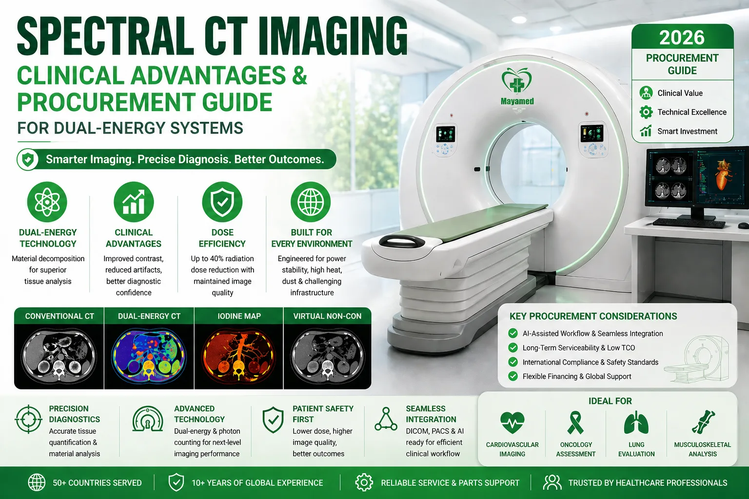
Unlike traditional single-energy systems, advanced ct imaging platforms leverage rapid kVp switching or dual-source architectures to capture attenuation data across multiple energy spectra. This capability enables precise material decomposition, allowing radiologists to isolate iodine contrast, quantify tissue composition, and generate virtual monoenergetic images that significantly reduce beam-hardening artifacts.
The clinical advantages of dual-energy ct benefits extend well beyond artifact reduction. By improving contrast-to-noise ratios and enabling quantitative tissue analysis, these systems enhance diagnostic confidence in oncology, cardiovascular, and musculoskeletal applications. Recent platform iterations also integrate photon counting ct detector technology, which eliminates electronic readout noise and improves Detective Quantum Efficiency (DQE) by up to 30%. The result is sub-millimeter spatial resolution (<0.3 mm), sharper tissue boundaries, and the ability to maintain diagnostic image quality while reducing patient radiation exposure by 20–40%.
Strategic Procurement & Deployment Considerations
Integrating next-generation CT into a clinical workflow requires aligning technical capabilities with institutional priorities. Procurement teams and hospital administrators should evaluate systems through the lens of long-term clinical utility and operational sustainability.
Workflow Integration & AI-Assisted Diagnostics
Modern scanners now embed machine learning algorithms for automated organ segmentation, real-time dose modulation, and iterative reconstruction. Prioritize open DICOM architectures that integrate seamlessly with existing PACS, RIS, and third-party AI workstations to avoid vendor lock-in and streamline radiologist reporting times.Maintenance & Quality Assurance Protocols
Preventive maintenance must be proactive. Establish structured QA routines—daily air/phantom calibrations, monthly tube warm-up cycles, and quarterly detector uniformity checks—to ensure consistent Hounsfield unit accuracy and extend gantry lifespan. Automated self-diagnostics can flag tube degradation or cooling inefficiencies before they impact clinical throughput.Addressing Common Deployment Hurdles
- Capital Allocation: High upfront costs can be mitigated through structured leasing, vendor-managed service agreements, or phased modular upgrades that scale with patient volume.
- Footprint Constraints: Compact 64- to 128-slice configurations now deliver spectral capabilities in standard room dimensions (~25–30 m²), eliminating the need for costly structural reinforcement or RF shielding modifications.
- Clinical Training & Retention: Partner with manufacturers that provide on-site application training, protocol optimization workshops, and continuous e-learning modules. Reducing the technologist learning curve directly impacts scan consistency and departmental efficiency.
Technical Specifications & International Compliance Standards
When evaluating system architecture, procurement specifications should move beyond basic slice counts and focus on measurable performance metrics. High-performance platforms typically feature:
- Detector & Gantry Performance: 128–256 detector rows, gantry rotation speeds ≤0.28–0.35s, and AI-accelerated reconstruction times <0.5s for routine clinical protocols.
- Dose Management: Adaptive statistical iterative reconstruction (ASIR/AIDR) paired with automated exposure control (AEC) and real-time dose tracking compliant with IEC 60601-2-44 safety standards.
- Regulatory Certification: Full compliance with ISO 13485 quality management systems, CE marking under EU MDR, and FDA 510(k) clearance. Reputable manufacturers provide complete technical dossiers, including electrical safety reports, EMC test certificates, and localized language manuals to accelerate regional health authority registrations.
Engineered for Challenging Environments: Infrastructure Adaptation for Emerging Markets
Medical imaging deployments across Sub-Saharan Africa, Latin America, and the Middle East face unique infrastructural realities. Grid instability, extreme ambient temperatures, and high particulate environments demand equipment engineered for resilience, not just peak laboratory performance.
- Power Grid Variability: Wide-input power supplies (100–240V AC, 50/60Hz) with integrated line conditioners, active power factor correction, and surge suppression protect sensitive detector electronics from voltage fluctuations and frequency drift common in regions with unstable municipal grids.
- Thermal Management: High-capacity, closed-loop liquid cooling systems paired with redundant HVAC interfaces maintain optimal tube and detector temperatures even in environments consistently exceeding 40°C (104°F), preventing thermal throttling during high-volume scan days.
- Environmental Sealing: IP54-rated gantry enclosures, anti-static filtration, and conformal-coated circuit boards mitigate dust ingress and humidity-related corrosion—critical for facilities in arid desert climates or coastal regions.
- Service Logistics & Uptime: Modular component design enables rapid field replacement by local biomedical teams. Combined with remote diagnostic telemetry, facilities can resolve software faults or replace consumables without waiting for international technician dispatch.
Conclusion & Next Steps
Upgrading to a spectral CT platform is a strategic commitment to diagnostic precision, patient safety, and operational efficiency. By aligning clinical requirements with robust technical specifications, regulatory compliance, and infrastructure-ready engineering, healthcare networks can secure imaging solutions that deliver measurable ROI across diverse practice environments.
With over 10 years of international export experience and a verified operational footprint across 50+ countries, Mayamed bridges the gap between cutting-edge imaging technology and practical, budget-conscious procurement. We don’t just distribute equipment—we deliver turnkey imaging solutions engineered for your facility’s clinical workflow and local infrastructure.
🚀 Ready to Evaluate Spectral CT Options for Your Facility?
- Request a detailed technical specification sheet & room planning guide
- Schedule a complimentary consultation with our biomedical engineering team
- Receive transparent pricing, flexible financing options, and global logistics support