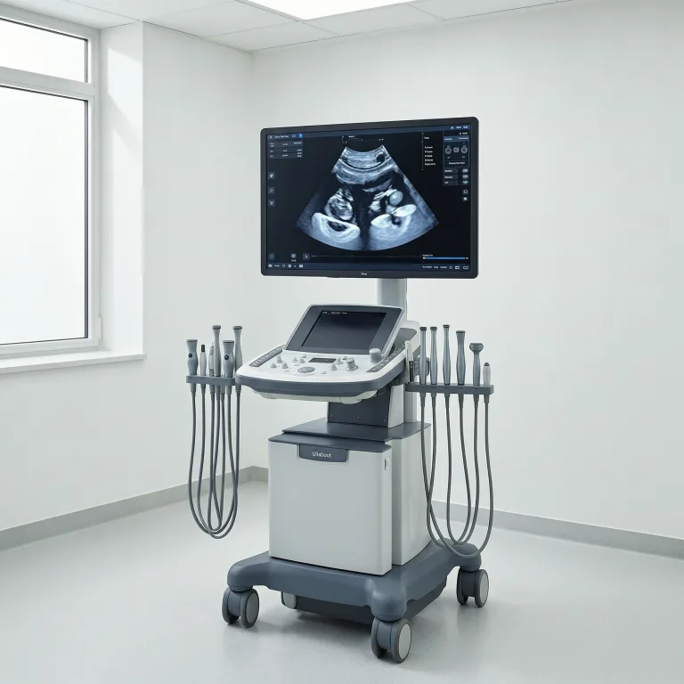
Understanding MRI Scans: Benefits, Prep & What to Expect
Magnetic Resonance Imaging (MRI) is a powerful, non-invasive diagnostic tool that creates high-resolution pictures of your body’s internal structures. Unlike X-rays or CT scans, MRI uses strong magnetic fields and radio waves—no ionizing radiation involved—making it especially safe for repeated use.
Key Benefits of MRI Scans
- Exceptional clarity for soft tissues like the brain, spinal cord, muscles, ligaments, and organs.
- Multi-directional views and advanced techniques (e.g., functional or vascular imaging).
- No radiation risk, ideal for children, pregnant patients (when benefits outweigh risks), and anyone needing frequent monitoring.
Compared to CT scans, MRI excels at soft tissue detail, while CT is faster (often 5-15 minutes) and better for bones or emergencies—but involves low radiation.
Common Reasons Doctors Order an MRI
Physicians frequently recommend MRI for:
- Brain and nervous system issues (tumors, stroke, multiple sclerosis)
- Joint, muscle, or spine injuries
- Abdominal and pelvic organ evaluation
- Blood vessel studies (MRA)
How to Prepare for Your MRI Scan
- Remove all metal objects (jewelry, watches, hairpins, clothing with zippers).
- Inform staff about any implants, devices, or metal fragments (e.g., pacemakers, cochlear implants, aneurysm clips).
- Wear comfortable clothes; you’ll likely change into a hospital gown.
- Eat and drink normally unless told otherwise (some contrast exams have restrictions).
- Arrive early to complete a detailed screening questionnaire.
Absolute Contraindications — MRI may not be possible if you have certain non-MRI-safe devices. Always consult your doctor.
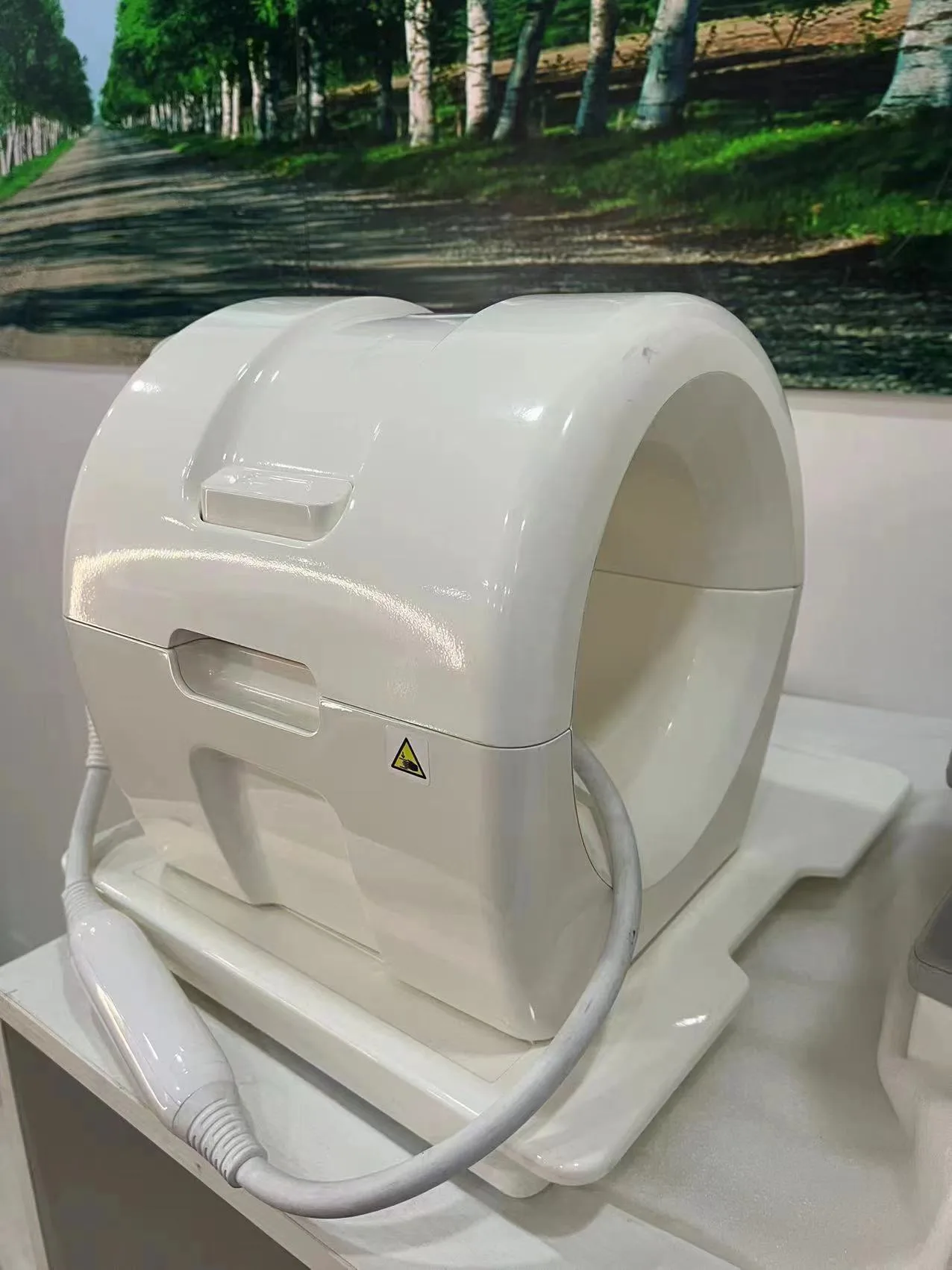
What Happens During the MRI Procedure
- Lie on a sliding table that moves into a tube-shaped scanner (open or wide-bore options available for comfort).
- The machine produces loud knocking sounds—earplugs or headphones are provided.
- Stay still for 20-60 minutes (scan time varies by area).
- For contrast-enhanced MRI, a safe gadolinium agent may be injected via IV; drink plenty of water afterward.
The exam is painless, though some feel anxious in enclosed spaces—sedation options exist for claustrophobia or children.
After Your MRI Scan
No recovery time is needed; resume normal activities immediately. Results typically take 1-3 days (longer for complex cases). Keep your images/disc and discuss findings with your referring physician.
Final Safety Tips for a Smooth MRI Experience
- Be completely honest about medical history and implants.
- Relax and follow technician instructions—movement can blur images.
- Modern scanners are quieter and more spacious than older models.
MRI remains one of the safest and most informative imaging methods available today. Always follow your healthcare provider’s personalized guidance.