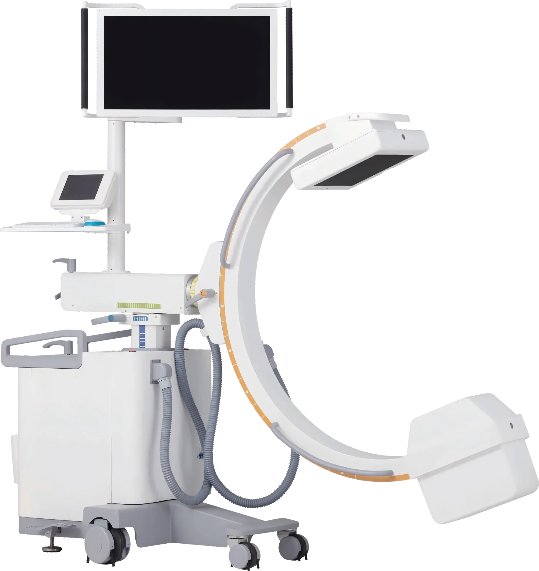
3D C‑Arm Imaging: Advanced Intraoperative X‑Ray Solutions
Modern 3D C‑arm systems combine rotational X‑ray acquisition with volumetric reconstruction to provide surgeons with real‑time three‑dimensional views during procedures. Proper setup, radiation safeguards, and artifact management are essential to get reliable, actionable intraoperative imaging.
How 3D C‑Arm Imaging Works
During a rotational sweep, the C‑arm acquires a series of 2D projections while the detector and X‑ray source circle the patient. Advanced reconstruction algorithms convert these projections into axial, coronal, and sagittal volumes for intraoperative review. Typical clinical acquisition spans a wide angular range to reduce artifacts and improve resolution.
Key Benefits Over 2D Fluoroscopy
- Immediate 3D confirmation of implant placement and bony anatomy.
- Better detection of malpositioned screws and fragment displacement.
- Enables minimally invasive approaches by reducing exploratory exposure.
- Supports navigation systems and robotic guidance when calibrated properly.
Practical Intraoperative Workflow
Follow a structured process to maximize image quality and safety:
- Pre-scan: calibrate the unit, confirm detector alignment, and position the patient for unobstructed rotation.
- Acquisition: perform a rotational sweep (wide angular coverage improves reconstruction). For thoracic-level imaging consider controlled apnea or short breath-hold to minimize motion artifacts.
- Post-acquisition: review orthogonal planes, enable artifact suppression, and acquire targeted additional scans if needed.
Metal Artifacts & Reduction
Implanted metals cause streaks and distortions. Modern systems include metal‑artifact reduction algorithms that use projection correction and iterative reconstruction to limit streaks. Best practices include slight angular offsets to avoid overlapping metal projections and enabling the system’s artifact reduction mode when available.
Radiation Protection & Best Practices
Reduce exposure by using low-dose protocols, collimation, and pulsed fluoroscopy. All staff should wear lead aprons and thyroid shields; distance and shielding remain effective. For pediatric and repeated procedures prioritize dose-sparing reconstruction settings and monitor cumulative dose.
Interoperability with Navigation & Robotics
When interfacing with navigation systems or surgical robots, lock the C‑arm after registration and verify the transform. Keep mechanical paths clear to avoid collisions. A stable, validated dataset improves guidance accuracy and reduces intraoperative surprises.