USD
USD EUR
EUR GBP
GBP CAD
CAD AUD
AUD HKD
HKD JPY
JPY BRL
BRL KRW
KRW CNY
CNY SAR
SAR SGD
SGD NZD
NZD INR
INR AED
AED MOP
MOP
- Description
- Download
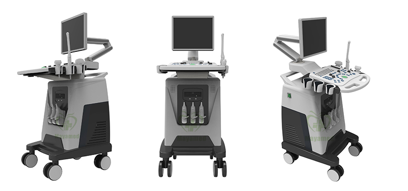
Specifications of MY-A028C Trolley Color Doppler
Windows 7 embedded operating platform, solid-state hard drive data storage,safe and reliable and boot quickly
| Display | 17 inch LED medical display |
| Probe connector | 3 |
| Operation interface | English (more languages as option) |
| Display mode | B、B+B、4B、B+M、M、C、PW、4D、B/C、B/C/PW、B/PW |
| Focus | 4 segments |
| Body mark | ≥57 |
| Image processing | Up/Down, Left/Right, Angular variation, Reverse |
| Measurement | Distance, circumference, area, volume, heart rate, diameter stenosis rate, area stenosis rate, angle, rhythm, speed. |
| Application | Abdomen,cardiac,skeletal muscle, early obstetrics, middle and late obstetrics, pelvic (uterine attachment) , small parts, urology, peripheral blood vessels, GW&EDD, fetal weight. |
| Character display | Date, time, name, sex, age, doctor, hospital, annotation (full screen character editing) |
| Cine loop | ≥ 1200 frames, continuous playback or view one by one. |
| Storage | Probe parameter, cine loop, measuring result, report can be saved and transferred to external storage device |
| Gray scale | 256 levels |
| Puncture guide | Available |
| Gravel positioning | Available |
| Dynamic range | 0-270dB |
| TGC control | 8 segments |
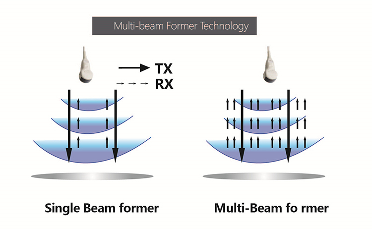
| Pre-processing | Variable aperture, dynamic apodization, dynamic digital filtering, multi-beam parallel processing technology, THI and so on. |
| Post-processing | Dynamic range 0-270DB, black and white afterglow 0-7, smoothing 0-7, gray curve 1-16, frame correlation, SHI, sound power, wall filter, cumulative number, baseline adjustment, sampling frame adjustment , spectral sampling volume, spectral sampling volume angle, the PRF (pu lse repetition frequency) and so on. |
| Blind zone | ≤4mm |
| Max display depth | 320mm |
| Geometric accuracy | horizontal ≤ 5%, vertical ≤ 5% |
| Resolution | lateral ≤ 2mm, axial ≤ 1mm |
| External interface | Video, USB, DICOM 3.0 |
| Display magnification | 16 kinds |
| Frame rate | 5-1016fps |
| Scanning range | 5% - 100% |
| Gain adjustment | Overall gain: 0 ~ 100%, PW gain: 0-15, CFM gain: 0-15 |
| Image optimization | 6 level adjustable |
| Smoothing | 8 level adjustable |
| Edge enhancement | 8 level adjustable |
| PRF | 16 level adjustable |
| Gray scale curve | 16 level adjustable |
| Acoustical power | 15 level adjustable |
| Hard Disc Storage | 120G |
Probe frequency range:
3.5MHZ convex probe: 2.0, 3.0, 3.5, 4.0, 5.5Mhz
6.5MHZ trans-vaginal Probe: 5.0, 6.0, 6.5, 7.5, 9.0Mhz
3.0MHZ Phased array probe: 2.0, 3.0, 3.5, 4.0, 5.0Mhz
7.5MHZ High frequency Linear Probe: 6.0, 6.5, 7.5, 10.0, 12.0Mhz
Volume probe: 2.0, 3.0, 3.5, 4.0, 5.5Mhz
Micro-convex probe R11: 2.0, 3.0, 3.5, 4.0, 5.5, 6.5, 7.5, 10.0 Mhz
Micro-convex probe R20: 5.0, 6.0, 6.5, 7.5, 9.0Mhz
System Overview
Application
Abdomen / Obstetrics / Gynecology /
Urology / Andrology / Small Parts / Vascular /
Pediatrics / Musculoskeletal
Electrical Power
Voltage: 100 V~240 V
Power: DC12.8V 3A
Frequency: 50/60 Hz
Conditions
Operating
Temperature: 5°C~40°C
Humidity: ≤80%
Pressure: 700hPa~1060hPa
Storage
Temperature: -5°C~40°C
Humidity: ≤80%
Pressure: 500hPa~1060hPa
Connectivity/Media/Peripherals
Transducer Ports: 3
USB Ports: 2
Hard Disc: 60GB (SSD)
120G/256GB SSD (Optional)
Footswitch: USB
Ethernet Port: 2(100Mb/1000Mb)
External Display: VGA
HDMI
USB Printer
Digital Laser Printer
Digital B/W Thermal Printer
Cine/Image Memory
Cine Memory: 1200 frame
Cine Review Speed: 1-5
Cine Review Loop
Cine Capture Function
DICOM Connectivity
DICOM3.0 Compliant
Image Storage
Storage Format:
PNG, AVI, BMP, JPEG, DICOM
Export Video Format: AVI
Export Image Format:
PNG, JPEG, BMP, DICOM
USB Flash Drive
Technology
Panoramic Imaging Tech
All-digital signal processing Tech
Multibeam formation Tech
Speckle Reduction Tech
Tissue Harmonic Imaging Tech
Dynamic Tissue Optimization Tech
Duplex & Triplex Synchronous Display
Directional Power Doppler
Imaging Parameters Preset
General Performance
Digital Broadband: 12288 channels
Beam-former: Re-programmable
Transmit Voltage: Adjustable (15 steps)
Beam-former Frequency Range: 1~40 MHz
Pan/Zoom
Real-Time Image Zoom
Zoom Range: 100%~400%
Up/Down/Left/Right Inversion
Hardware Specification
LCD Monitor
Size (Diagonal): 15”
Contrast Ratio: 800:1
Resolution: 1024×768 pixels
Brightness: 230 cd/m2
Color Depth: 24bit
Rotate Angle: ± 90°
Grey Levels: 256
Embedded Speakers
Impedance: 4Ω
Power: 5 W
UPS (Optional)
Imaging Performance
Startup Time (Max):
Avg. < 90 seconds
Preset Switching Time:
Avg. < 1 second
Storage Time (Image to Disk):
Avg. < 0.5 second
Transducers
Convex Probe
Frequency: Central 3.5 MHz
Min. 2.0 MHz
Max. 5.0 MHz
Pitch: 0.516 mm
Radius: 60 mm
Number of Elements: 96
Linear Probe:
Frequency: Central 7.5 MHz
Min. 6.0 MHz
Max. 12.0 MHz
Pitch: 0.352 mm
Radius: N/A
Number of Elements: 96
Trans-vaginal Probe:
Frequency: Central 6.5 MHz
Min. 5.0 MHz
Max. 9.0 MHz
Pitch: 0.216 mm
Radius: 10 mm
Number of Elements: 96
User Interface
• Intuitive Windows-based operating principles
• User-centric control panel with HomeBase layout and control customization
• On/Off task light and back-lit illumination of control panel
• Variable brightness indicates active state of function keys
• Easily accessible, full size QWERTY keyboard for text entry, function keys and system programming
• Cine Playback,Multiple Arrows,Configurable Worksheets,Exam Review,Pictograms (Body Marks),System Setup Menu
Imaging Modes
B, 2B, 4B, M, B/M, B/C, B/D,
B/C/D, B/CFM/D, PDI
Color, Dual Color
Simultaneous 2D/Color Compound
PW, Duplex/Triplex
CFM, CDE, PD, Directional PD, CD
B-Mode
Maps: 17
Focus Count: Max. 4
Gain: 0~255
TGC: 8 Sliders
Dynamic Range: 0~150dB
Depth: Max. 30cm
Acoustic Power: 0 ~ 15
Chroma: 0~7
Grayscale Levels: 256
Frame Rate: Max.1028Hz
Persistence/ Frame Average: Up to 7
Image Optimization: 0~6
Top/Bottom/Left/Right Reverse
Speckle Reduction
Second Harmonics
M-Mode
Maps: 16
Chroma: 16
Sweep Speed: 3
Gain: 0~255
Distance: Point-to-Point
Max Steer Angle: 15°
Diameter Reduction: Point-to-Point
Heart Rate/Time/Distance/Slope
Side-by-Side/Bottom
Color Mode
Gain: 0~255
Color Maps: 0~6
Doppler Steer Angles: 7 Steps
Wall Filter: 0~3
Spatial Filtering: 0~3
Acoustical Power: 0 ~ 15
Doppler Multi-Frequency: 2MHz~10MHz
Line Density Max: 256 Lines
Persistence/Frame Average: 0~7
Bloodstream Gain: 0~127
Packet Size: 8~15
Baseline/Invert/Color ROI/M-Mode
PW Mode
Gain: 0~255
D Linear Velocity: 0~2
Doppler Steer Angles: 7 Steps
Edge Enhancement: 0~7
Wall Filtering: 0~3
Audio: 0~255
Correction Angle: 80°/-80°
Doppler Multi-Frequency: 2MHz~10MHz
Pulse Repetition Frequency: 2khz~6khz
Chroma: 0~7
Baseline Adjustment: 0~6
Three Synchronizations
Spectrum Inversion
Measurement Specifications
Analysis Packages
Basic
Obstetrics
Gynecology
Urology
Andrology
Peripheral Vascular
Venous
Small Parts
Orthopedic
Basic Measurement
B-mode basic measurement:Distance, Angle, Perimeter and Area (ellipse or track-based method), Volume, Histogram, Sectional Map
M-mode basic measurement:Heart Rate, Time, Distance, Speed
Obstetrics
All general measurements and calculations
Obstetrical data versions to calculate gestational age:
— Two fixed data revisions: Asia & Europe
— One editable data version: User Custom
— Each version can estimate the gestational age and expected date of confinement based on measured gestational sac (GS), biparietal diameter (BPD), crown-rump length (CRL), femur length (FL), humeral length (HL), abdominal transverse diameter (ATD), vertebral length (LV), occipitofrontal diameter (OFD), abdominal circumference (AC) and head circumference (HC)
Obstetrical Report
Amniotic Fluid Index (AFI)
BPD/OFD, FL/AC, FL/BPD and HC/AC ratio
Estimate Weight Of Fetus
Gestational Age
Expected Date Of Confinement (LMP/BBT)
Fetal Biophysical Score
Fetus Growth Curve
Gynecology
All general measurements and calculations
Uterus, Ovary, Follicle
Gynecology Report
Urology
All general measurements and calculations
Kidney, Bladder, Residual Urine Volume
Urology Report
Andrology
All general measurements and calculations
Prostate, Testis
Prostate Specific Antigen (PSA)
Prostate Specific Antigen Density (PSAD)
Andrology report
Small Parts
All general measurements and calculations
Thyroid, Mammary Gland, Nodule
Small Parts Report
Peripheral Vascular
All general measurements and calculations
Area Stenosis
Vessel Diameter Stenosis
Peripheral Vascular Report
Venous
All general measurements and calculations
Right and left extremity measurements
Venous patient report
Orthopedic
All general measurements and calculations
Right and left hip angle measurement
Hip angle patient report
Optional:
SONY Viedo printer 898MD
Mitsubishi Viedo printer P93w-Z
You May Like

-
$38,000.00

-
$1,700.00

-
$11,300.00
")
-
$6,000.00

-
$8,680.00

-
$12,500.00

-
$15,500.00

-
$3,000.00

-
$1,451.00